|
Modalities
- Physical Examination and Specific Clinical Tests
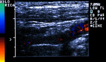
- Color Dopler Ultrasonography
(Dublex USG)
- venography
Diagnosing
Venous Disease and Pulmonary Embolism
One or more of the
following techniques are used for diagnosis of venous diseases
Duplex or Doppler Ultrasound – This non-invasive technique is used to “see” clots and
dilatations in the blood vessels and demonstrate venous flow
direction.
Venography
- a thin, flexible catheter, is inserted into the blood vessels. A
local anesthetic is given to numb the skin where the catheter is
inserted, contrast agent, or dye, is injected through the catheter to
highlight the blood vessel.
Diagnosing Pulmonary Embolism
A V/Q scan (sometimes called a V/P or ventilation/perfusion scan) is a
nuclear medicine test in which short-acting radioactive particles are
injected through a vein or breathed into the lungs. If there are areas
of the lung that do not “take up” the particles, it is an indication
that there may be a blood clot.
Cafer Abbasoğlu
jan 2007
|